RENCI-enhanced software helps reduce false positives and maintain compliance.
Currently, about 12 percent of the female population will develop invasive breast cancer in their lifetimes, and for women in the U.S., breast cancer death rates are higher than those for any other cancer, besides lung cancer, according to the American Cancer Society. Despite these harsh realities, the death rates from breast cancer have steadily decreased since 1989, a trend often attributed to treatment advances and increased screening.
Research indicates that mammography accurately identifies about 78 percent of women who have breast cancer. In women over 50, that accuracy rises to about 83 percent. However, screening mammography also has limitations, including the potential to miss cancers (false negatives) and to call women back for additional work-up when no cancer is present (false positives).
Research groups such as the University of North Carolina at Chapel Hill’s Carolina Mammography Registry (CMR) work to improve breast cancer screening by partnering with breast imaging facilities to make data available to researchers. CMR works with practices across North Carolina to collect information from women and radiologists, which is then combined with cancer outcome data to provide feedback to radiologists on the quality of their mammography interpretations.
In addition to their work within the state, CMR is a member of the Breast Cancer Surveillance Consortium, a nationwide collaboration dedicated to conducting rigorous research to improve breast cancer detection. This research is used to inform national guideline development on how often and when women should have screening mammograms.
When Dr. Louise Henderson, CMR principal investigator and director, assumed leadership in 2011, one of her first moves was to upgrade the CMR Data System (CMDS), custom radiology information software that had been in use since the CMR started operations in 1994. Henderson reached out to developers at the Renaissance Computing Institute (RENCI) with a request to improve the existing CMDS system to be compatible with current operating systems and to increase the ability to capture additional data fields in a user-friendly format.

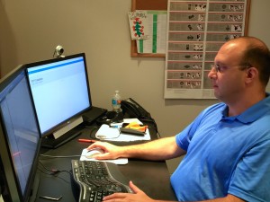
Oleg Kapeljushnik. software developer, helped to update the CMR data system.
The CMDS allows breast imaging facilities to collect patient risk factor and history data, breast imaging information, findings, and the radiologists’ interpretation as well as recommendations. These data are then used to generate reports and reminder letters, which are crucial to ensuring appropriate follow-up. Developers at RENCI have collaborated with the CMR to improve on these functionalities by adding new features related to systems compatibility, reporting, and compliance with changing laws.
“The way I see this software is as a way for practices to make actual patient data available to researchers. With this new version, practices are able to keep using the software on new platforms with added features and abilities that allow them to be more flexible in addressing changing needs and regulations,” said Oleg Kapeljushnik, software developer.
Using the updated system enhanced by RENCI, CMR-participating breast imaging facilities have a quick and effective way to incorporate additional data fields, making the collection of new imaging modalities or risk factor information straightforward.
For example, the use of digital breast tomosynthesis, a new type of mammography that has been referred to as 3D mammography has begun to spread across the state. With the redesign of CMDS by RENCI, breast imaging facilities that utilize CMDS are easily able to add a data field to specify that the breast imaging examination is a 3D mammogram. Not only is this valuable for the facility, but it is also important for the collection of data as researchers and practitioners strive to evaluate the effectiveness of new technologies for detecting breast cancer.
“Using the revamped CMDS software, the CMR-participating facilities are able to continue to collect breast imaging data that are vital to ongoing research efforts in breast cancer screening,” said Henderson.
Overall, providing radiologists with improved software to collect, access, and analyze data means that more radiologists can track and evaluate their results to improve the quality of care they give to their patients.


