Overview
Melanoma, the most dangerous form of skin cancer, is typically diagnosed by pathologists who visually examine biopsies from unusual skin growths. Although many pathologists are well-practiced at identifying melanoma, computer-based image analysis could help refine the diagnostic process by quantifying features identifying these cancerous tissues. In a project led by melanoma specialist Dr. Nancy Thomas, professor in the School of Medicine at UNC Chapel Hill, RENCI collaborates with researchers in computer science, statistics and operations research, dermatology, and pathology to apply image analysis approaches to aid in diagnosis and prognosis of melanomas from biopsy slides.
The Melanoma Image Analysis project aims to enhance prediction of patient survival, link melanoma phenotypes to specific genetic mutations, and guide future treatment options. RENCI experts work with the project team to develop image analysis algorithms for extracting melanoma features, such as stain intensity, density of cell nuclei, and nuclear area and shape, that can lead to clinically relevant tests.
Funding
University Cancer Research Fund
Collaborators
- Nancy Thomas, M.D., School of Medicine, UNC Chapel Hill
- Marc Niethammer, department of computer science, UNC Chapel Hill
- Steve Marron, department of statistics and operations research, UNC Chapel Hill
- John Woosley, M.D., department of pathology and laboratory medicine, UNC Chapel Hill
Project Team
- David Borland
- Charles Schmitt
Publications
Macenko, M., Niethammer, M., Marron, J.S., Borland, D., Woosley, J.T., Xiaojun Guan, Schmitt, C., Thomas, N.E. A method for normalizing histology slides for quantitative analysis. IEEE International Symposium on Biomedical Imaging: From Nano to Macro, 2009. ISBI ’09. 2009.
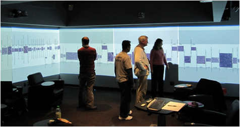
- The project team examining melanoma data in the Social Computing Room, one of RENCI’s facilities for visualizing large datasets.


